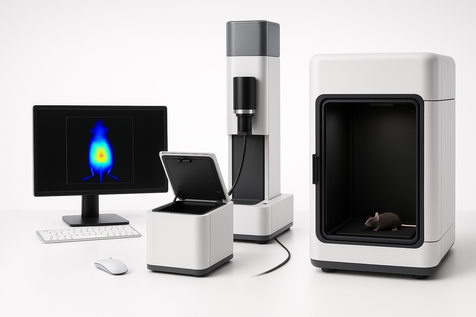
In-vivo bioluminescent imaging (BLI) has become one of the most widely used tools for studying biology in real time. At its core, the technique takes advantage of bioluminescence, the natural ability of organisms like fireflies, marine bacteria, and deep-sea shrimp to produce light through enzyme-driven chemical reactions. In research settings, scientists can introduce luciferase genes into cells, tissues, or pathogens of interest. When the substrate luciferin is supplied, the luciferase enzyme generates light that can be picked up externally by highly sensitive imaging cameras—turning invisible biological processes into something we can actually watch unfold inside a living animal.
What makes BLI so powerful is its ability to track changes over time in the same subject, without invasive procedures. This reduces variability, lowers the number of animals needed, and provides a much clearer picture of dynamic processes like tumor growth, infection spread, stem cell survival, or drug response. It has proven especially valuable in fields such as oncology, infectious disease, neuroscience, and regenerative medicine, where following events in real time can be the difference between guessing and knowing.
The technique’s strengths lie in its sensitivity, low background noise, and capacity to generate both spatial and temporal information. But BLI is not without limitations. Light signals are weakened as they pass through tissue, substrate delivery can be a bottleneck, and applications are generally restricted to small animal models. Even so, the method continues to evolve and remains one of the most insightful ways to study complex biology in vivo.
One of the most exciting recent advances is the development of infraluciferin, a red-shifted variant of luciferin. Unlike conventional luciferin, infraluciferin produces longer-wavelength light that travels through tissues with less scattering and absorption. This means clearer images, deeper penetration, and more reliable signal detection. In addition, infraluciferin provides stronger sensitivity, more stable signals, and, when used with engineered luciferases, the ability to generate multiple colors from the same system. These improvements open the door to multiplex imaging, allowing researchers to follow several biological processes simultaneously and to study tissue and disease morphologies that standard luciferin simply can’t reveal.
